
Identifying enteral feeding complications at home involves monitoring for warning signs such as tube blockages, site redness, leaking, or digestive distress like bloating and aspiration risks. Early recognition and proactive management through consistent hygiene and flushing protocols are essential for preventing hospital readmissions and ensuring patient safety.
Transitioning from hospital to home-based enteral feeding is a significant milestone, but it often brings a hidden layer of anxiety regarding potential complications. The responsibility of monitoring a PEG tube or nasogastric line can feel overwhelming when clinical staff are not immediately present. Early recognition of issues is not just about technical maintenance; it is the cornerstone of preventing hospital readmissions and ensuring long-term nutritional success within the Australian home care setting. In this guide, we provide practical expertise on identifying the early warning signs of aspiration, stoma site infections, and mechanical tube failures. You will also learn how to differentiate between minor gastrointestinal distress and serious malabsorption issues. By understanding these clinical markers and knowing when to engage registered nursing oversight, you can confidently manage enteral nutrition while maintaining the highest standards of safety and care in a domestic environment.
Managing Enteral Nutrition in an Australian Home Setting
For families across Norwest and Greater Sydney, transitioning from a hospital ward to Home Enteral Nutrition (HEN) represents a significant shift in care dynamics. While tube feeding is a life-sustaining intervention, the move to a domestic setting shifts the critical responsibility of monitoring from a 24/7 clinical team to family members and support workers. This transition can be daunting, as the quiet efficiency of a hospital is replaced by the realities of daily life.
The primary objective of this guide is to bridge the gap between initial hospital discharge training and the practicalities of long-term care. Spotting enteral feeding complications at home early is the most effective way to prevent emergency department presentations and ensure the patient remains stable and well-nourished. Early detection of issues like site irritation or minor blockages allows for proactive intervention rather than reactive crisis management.
Build Your Community (BYC) acts as a managed healthcare brokerage to support this process, providing clinical oversight through the SALUS clinical care pathway. By integrating professional nursing expertise with NDIS enteral feeding support, we ensure that caregivers have a clear framework for monitoring. Our goal is to empower families with the clinical confidence required to manage HEN safely and effectively within the community.
Spotting Aspiration: The Critical Warning Signs

Aspiration occurs when formula, saliva, or gastric contents enter the airway instead of the oesophagus. While overt aspiration is marked by obvious choking or gasping, silent aspiration is far more insidious. It often occurs without a clear cough reflex, particularly in individuals with neurological impairments. Because it lacks dramatic symptoms, it is frequently overlooked until it develops into aspiration pneumonia, making it one of the most serious enteral feeding complications at home.
Identifying aspiration requires caregivers to move beyond looking for obvious distress and instead focus on subtle auditory and physiological changes. These specific warning signs include:
A wet or gurgly quality to the person’s voice or breathing, sounding as if there is fluid sitting on the vocal cords.
Persistent, irritant coughing that occurs during the feed or up to thirty minutes after completion.
Sudden, unexplained breathlessness or a noticeable increase in the effort required to breathe.
A sustained drop in oxygen saturation levels for patients who utilise a pulse oximeter for monitoring.
Watery eyes or sudden skin flushing during the administration of the feed.
If any of these signs are detected, the most critical action is to stop the feed immediately. Leaving the pump running or continuing a gravity feed significantly increases the risk of lung injury. Once the feed is paused, you must contact a clinical professional to assess the patient’s chest sounds and swallow safety.
Through the SALUS clinical care pathway, BYC provides the necessary oversight to manage these risks. As a managed healthcare brokerage, we ensure that your NDIS enteral feeding support includes a robust escalation plan. This clinical framework ensures that when subtle signs of aspiration appear, they are met with a professional nursing response that evaluates the feeding position, delivery rate, and formula consistency to prevent a recurrence.
Identifying PEG Stoma Site Infections and Skin Issues
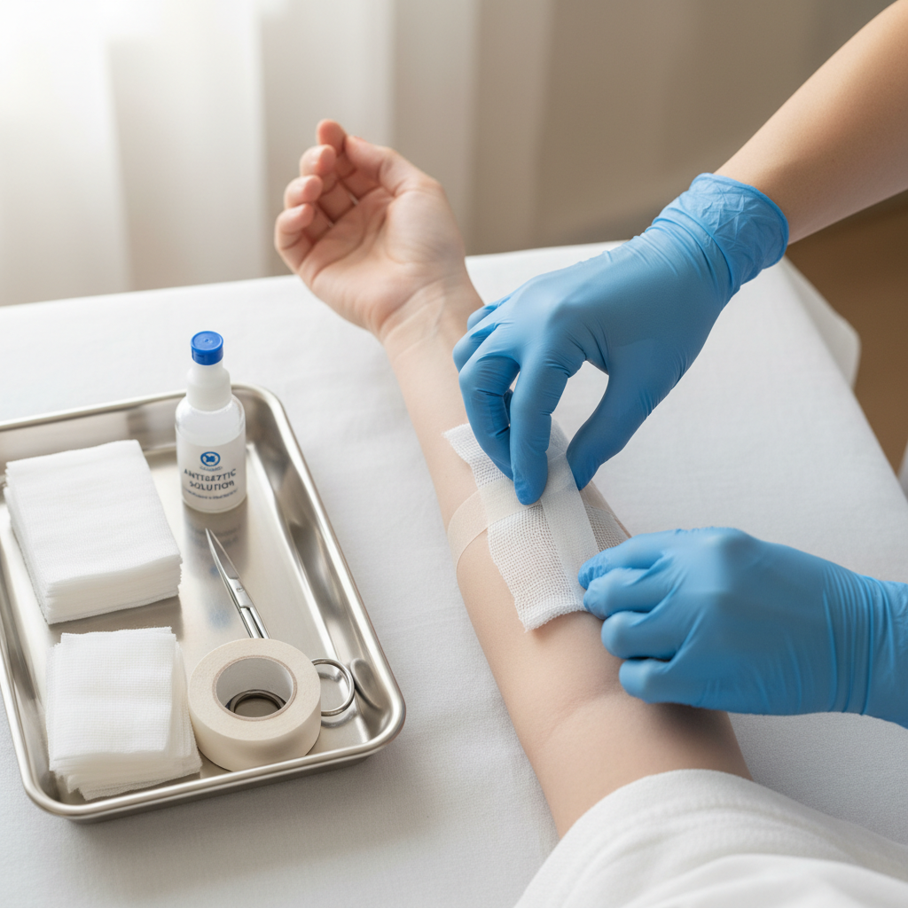
Monitoring the stoma, which is the entry point where the Percutaneous Endoscopic Gastrostomy (PEG) tube meets the skin, is a daily requirement for preventing enteral feeding complications at home. It is common to see a small amount of clear or pale yellow fluid, known as serous discharge, around a healthy site. This is part of the body’s natural healing process and is generally not a cause for concern. However, caregivers must be able to distinguish this from the specific symptoms of an infected PEG site.
Clinical indicators of a localized infection typically include: - Thick, opaque discharge that appears green, white, or dark yellow. - A foul or unusual odour emanating from the stoma area. - Spreading redness, heat, or significant swelling of the skin surrounding the tube. - Persistent pain or tenderness when the tube is gently touched or rotated.
In the humid Australian climate, particularly during the summer months in Greater Sydney and the Norwest region, moisture trapped under the external bolster can rapidly lead to skin maceration. Maintaining a clean, dry environment around the stoma is the primary defence against fungal and bacterial growth. Families may also observe "granulation tissue," which appears as red, moist, or slightly bumpy flesh at the tube entry. While this often looks alarming to the untrained eye, it is simply an overgrowth of healing tissue and small blood vessels. It is manageable but requires professional assessment to ensure it does not interfere with the tube’s position or cause bleeding.
A leaking PEG stoma presents a different risk, as gastric contents are highly acidic and corrosive to the skin. If formula or stomach acid escapes around the site, it can cause rapid skin breakdown and painful excoriation. As a managed healthcare brokerage, BYC ensures that NDIS enteral feeding support includes proactive site reviews. Through the SALUS clinical care pathway, we facilitate Registered Nursing oversight to manage bolster tension and skin integrity, ensuring minor irritations are resolved before they compromise the patient’s health.
How to Tell if a Feeding Tube is Blocked or Displaced
Mechanical complications are common but manageable if identified early. A blockage often manifests as sudden, tactile resistance when attempting a routine water flush. If you are using an electronic pump, it may trigger occlusion or high pressure alarms repeatedly, even after you have checked for external kinks. You might also notice visible residue, clumping, or darkened patches of old formula within the tube walls. One of the leading causes of these enteral feeding complications at home is the administration of improperly crushed medications. Pills that are not ground into a fine, uniform powder or adequately dissolved in water can easily lodge in the narrow lumen of the tube, creating a physical barrier that stops the flow of nutrition.
Tube displacement requires a different set of observations. For many patients, the tube is marked at the exit point with centimetre increments. Caregivers should record the measurement at the skin level during every dressing change to establish a baseline. A change in this number indicates that the tube has migrated either further into the stomach or partially out of the stoma. Beyond the markings, internal displacement can cause localized discomfort, sharp pain during feeding, or a feeling of fullness that was not previously present.
If a feeding tube is completely dislodged and comes out, it is a medical emergency. The stoma tract can begin to close in as little as two to four hours, making reinsertion difficult or impossible without surgical intervention. In these instances, cover the site with a clean dressing and seek immediate hospital care. As a managed healthcare brokerage, BYC integrates NDIS enteral feeding support with the SALUS clinical care pathway to provide families with clear protocols. This clinical oversight ensures that support workers and family members know exactly how to monitor tube position and manage flushes to prevent these mechanical failures.
Recognising Poor Absorption and Gastrointestinal Distress
While mechanical issues are visible, physiological enteral feeding complications at home are often detected through changes in the patient’s digestion and comfort. To answer a common question for families; what are two signs that the patient is not absorbing their enteral feed? The most frequent indicators are high gastric residuals, often manifesting as a feeling of excessive fullness or vomiting shortly after a feed begins, and persistent diarrhoea.
These symptoms are not merely expected side effects of tube feeding. Instead, they indicate that the gastrointestinal tract is struggling to process the volume, rate, or concentration of the formula. Severe abdominal bloating and distension are further signals that the feed may be fermenting in the gut rather than being absorbed. When these signs are ignored, the risk of dehydration and malnutrition increases significantly; these are leading causes of avoidable hospital readmissions for HEN patients across NSW.
A clinician must review the feeding regimen if these symptoms persist. This might involve adjusting the delivery rate on the pump, changing the osmolarity of the formula, or introducing a different feeding schedule. As a managed healthcare brokerage, BYC facilitates this clinical review process. Through the SALUS clinical care pathway, we ensure that NDIS enteral feeding support includes regular monitoring of tolerance. By identifying poor absorption early, we can coordinate with dietitians and nurses to recalibrate the nutrition plan, ensuring the patient receives the caloric intake they need without the distress of gastrointestinal failure.
When to Escalate: The Role of Registered Nursing Oversight

Distinguishing between daily monitoring and clinical assessment is vital for maintaining safety in a home environment. While support workers are skilled in the routine administration of formula and basic site hygiene, they operate within a defined scope that does not include clinical diagnosis. When subtle changes in tolerance or site health occur, the expertise of a Registered Nurse is required to determine if the care plan needs immediate adjustment. This professional oversight prevents minor issues from escalating into systemic failures.
Certain symptoms demand an immediate transition from home care to emergency medical intervention. Peritonitis, a serious inflammation of the abdominal lining, can occur if gastric contents leak into the peritoneal cavity. Caregivers must be vigilant for red flag symptoms, including a high fever, a board-like rigidity of the abdomen, and intense, localized pain that worsens with movement or touch. These signs indicate a critical failure and represent the most severe enteral feeding complications at home. In such cases, the priority is rapid hospitalisation rather than waiting for a scheduled nursing visit.
Build Your Community (BYC) functions as a managed healthcare brokerage to ensure this escalation process is never left to chance. By implementing the SALUS clinical care pathway, we provide a framework where clinical professionals actively manage the engagement. This ensures that NDIS enteral feeding support is underpinned by qualified nursing oversight, allowing for the early detection of deteriorating health. Our approach moves care beyond simple task completion, placing a clinical safety net under every participant to ensure that emergency signs are recognised and acted upon before they become life-threatening.